


Table of Contents
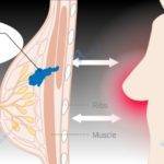
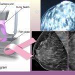
Ultrasound is one of the most frequently used modalities or machines in the screening and diagnosis of breast cancer.
After mammography, an ultrasound could be ordered as a next-step follow-up measure if the mammogram showed suspicious abnormalities, you had complaints suggestive of interval breast cancer, you have very dense breasts, or at high risk of cancer, among a few other factors. Ultrasound helps to distinguish a cyst (fluid-filled sacs or cavities) from a tumor.
Interval breast cancers are cancers that develop between your mammogram screening schedules and are usually a cause for concern. They are more aggressive and define more than a fifth of all breast cancers according to the Oregon Health and Science University.
Women usually freak out when they get a call-back following their mammograms but there is no reason for this as call-backs often turn out negative. They are to ensure your safety and good health.
The American Cancer Society (ACS) has opined that less than 10% of all call-backs turn out positive for breast cancer.
In this article, we’ll take a look at why you do an ultrasound after a mammogram.
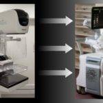
How it works.

The ultrasound machine uses a probe called a transducer to send very high-frequency sound waves into your body. So high that they are inaudible. This probe also acts as a receiver to the same sound waves and converts them into electrical signals via special objects called quartz crystals.
These sound waves travel into your breast till they reach the boundary of internal structures. At this point, some of the waves are reflected to the transducer (probe) as echoes while others carry on with their journey to other boundaries.
The electrical impulses from the received echos are sent to a central processing unit which converts them into an image that displays on a monitor in real-time.
The shape of the echoes received will depend on the curvature of the boundaries encountered. The intensity is also determined by the absorption properties of the internal structure, to sound waves.
Fluids absorb more waves thereby reflecting fainter or weaker echoes back to the transducer which makes them appear dark in the monitor as are tumors.
Fibroglandular (fibrous and glandular) structures in your breast, on the other hand, appear lighter in monitors, unlike tumors. This is exactly what makes ultrasound very useful if you have dense breasts.
Reasons for breast Ultrasound.
Ultrasonography complements mammography for breast screening and diagnostic purposes. It’s often the next-step approach in investigating abnormalities found in a mammogram
to know if it’s a cyst or a solid lump.
It’s also capable of detecting things that may be missed in regular mammography like superficial growths and when the breasts are dense. Additionally, it’s also an excellent tool in needle biopsies as it enables radiologists to see the location of a tumor in real time.
9 reasons you do an ultrasound after a mammogram.
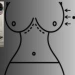
1. Abnormal Mammogram.

Image credit: Radiology Assistant
For as long as your regular screening shows there are no problems, you will only have to go home till your next appointment for a screening mammography. However, if on any of the occasions, an abnormality is detected, a call-back is certain.
This will often mean doing an ultrasound for further investigation.

Image credit: Radiology Assistant
For pre-menopausal women (mostly those below the age of 45), ultrasonograms happen to be more accurate and clearer than regular mammograms because of dense breast tissues.
This is because, in mammograms, both the fibroglandular tissues responsible for dense breasts and tumors appear white thereby making it difficult to differentiate the two.
Ultrasound, on the other hand, shows tumors to be dark and fibroglandular tissues as lighter in color. This makes it much more accurate in the diagnosis of cancer in younger women.
Most cancers, however, occur in older women but when they are found in young women they are often more aggressive and difficult to deal with.
A lot of women who show up for mammograms have dense breasts. About half of all women are believed to have dense breasts.
3. Complaints. If in your scheduled screening mammogram appointment you make complaints that suggest a problem, you may be recommended to have a diagnostic mammogram instead and an ultrasound afterward.
This is to ensure nothing is missed if there is anything at all.
4. Age. The older you are, the fattier your breasts (though not in every woman), therefore the more accurate will be the mammography. On the contrary, the younger you are, the less fatty and by extension the denser your breasts tend to be. This still has to do with breast density.
For this reason, ultrasound is more accurate and may be used to complement your mammogram if you are young and your doctor thinks it’s necessary.
5. Being at high risk. If risk assessment tools place you at high risk of developing breast cancer, you may have to start your annual mammogram screening earlier than usual by which time your breasts could be significantly dense.
This could mean having you do an ultrasound test in addition to mammography as part of your regular screening.
You are considered to be at high risk if;
- Test results show you have the BRCA1 and BRCA2 gene mutations.
- Risk assessment tools place your risk level at 20% to 25%.
- You, for any reason, have had radiation therapy between the ages of 10 – 30 years.
- Any of your first-degree relatives (parents or siblings, for instance) is known to have the genetic mutation mentioned above but you haven’t yet done the test.
- You or any first-degree relative is known to have had certain syndromes like Cowden or Li-Fraumeni, etc.
6. Unclear mammogram result. When your mammogram gives inconclusive results it may be a reason for an ultrasound to be ordered for clarity.
7. First-ever mammogram. If you report for your first-ever mammogram there will be no previous report to use for comparison, this may serve as a reason to have an ultrasound done after the mammogram especially when something shows up that needs more investigation.
8. Unavailability of previous mammogram results. Reporting to a new mammogram screening center without any previous report is a little different from having your first-ever mammogram. Just as in point 7 above, you may end up doing an ultrasound after the mammogram.
According to the American Cancer Society (ACS), it’s also common if you are pre-menopausal.
If you have any of the BRCA1 or BRCA2 gene mutations, informing your family members so they understand their own risk of cancer may be life-saving. If you don’t know how to go about this, use this tool to practice the best way of getting them properly informed as recommended by the Centers for Disease Control and Prevention (CDC).
9. Presence of perfumes, deodorants, and powders: You are advised not to apply these on your mammogram day or clean them up from around the breasts before your mammogram. These substances appear on your mammogram as abnormalities which can lead to a call for ultrasound for confirmation.
When Ultrasound hurts.
Ultrasonography is meant to be a painless procedure but in very few cases, it may bring about pain and this may be due to an underlying non-cancerous cyst.
If for any reason you are having pain before the procedure, like post-breast trauma (that is after breast trauma) or a recent biopsy that hasn’t healed, it shouldn’t come as a surprise if you feel pain during an ultrasound exam.
Mammography, on the other hand, is known to cause discomfort and in some cases, pain.
Why an abnormal mammogram but a normal Ultrasound?
Though Ultrasound has proven to be an invaluable modality in breast cancer screening and diagnosis, particularly in younger women, it may still miss things visible in a mammogram. In such instances, it results in a false negative report.
Situations that could give rise to this include:
These are tiny specks of calcium deposits in the breast which are usually visible in a mammogram but missed in an ultrasound. Unfortunately, calcifications may turn out to be the only sign of cancer sometimes.
2. Large breasts. Ultrasound is known to be much less accurate if you have large breasts. Mammography, on the other hand, has no problems giving clearer images even in large breasts unless the compression wasn’t adequate.
Inadequate breast tissue compression during a mammogram may lead to the superimposition of structures within the breast. This often results in having a repeat mammogram for clearer images.
Having large breasts is one reason why an ultrasound may show normalcy whereas a mammogram has shown an alarming development.
3. Deep-seated abnormalities. Whereas ultrasound is good at detecting superficial growths in a breast, it could miss deeper abnormalities within the breast. This invariably could result in a normal ultrasonogram but an abnormal mammogram.
How long does it take to get your results?
Where everything went normally without hitches, ultrasonography will take about 15 to 30 minutes and you could be informed of your result after the test, before leaving the health or diagnostic facility.
Notwithstanding, it takes a day or two for the report to be ready and sent to your doctor.
I hope this was helpful. If it wasn’t, do tell me the areas you are concerned about and also how I can serve you better.
You may also want to read about mammograms for breast cancer and Recommended age for mammograms.


I almost never leave a response, but I did some searching and wound up here: Explicit reasons
you do an ultrasound after a mammogram. And I actually do have
a couple of questions for you if you tend not to mind.
😛 and, if you are writing at additional sites,
I would like to keep up with everything new you
have to post. Could you list the complete
urls of your public pages like your Facebook page, twitter feed, or linkedin profile?
Hello Venus,
Everyone is free to drop a question or leave a comment.
That’s what this section is meant for.
The links to our various social media profiles are provided at the footer section here on our website and visually represented by icons. Clicking on an icon of your choice will take you there.
Very relevant.
Very important piece.
The most relevant.
Excellent, what a web site it is! This weblog gives
helpful information to us, keep it up.
I like the helpful information you provide in your articles.
I will bookmark your weblog and check again here frequently.
I am quite certain I’ll learn a lot of new stuff right here!
Best of luck for the next!
I simply stumbled upon your website and I did in fact enjoy your weblog posts. I will be subscribing to your feeds.
Thanks for sharing such know-how, it’s good to read
this weblog, and I pay a quick visit to this weblog everyday.
Excellent work from a gifted writer. You know this just too well.
Just want to say your article is amazing. Thanks a million and please keep up the great work.