


Table of Contents
If you have ever had a mammogram that reveals breast cancer you may already know what it looks like. However, for the vast majority of women who will never come down with the disease, it is imperative to give them ideas of what radiologists look for in mammograms conducted for breast cancer.
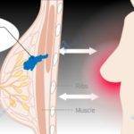
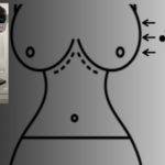
I will start with the anatomy of some of the important parts of a breast before proceeding with the mammogram of breast cancer.
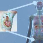
Breast Anatomy.

• Lactiferous Ducts: These are tube-like structures linking the nipples with the lobules where milk is produced. I guess you already know they help in transporting milk from the points of production to the end of consumption.
• Fibrous tissues and Fats (also known as adipose tissues): These help hold all other structures together and are chiefly responsible for the shape and size of your breasts. The adipose tissues, however, go the extra mile to aid the development of breast cancers as well.
This, they achieved partly by secreting growth factors the cancer cells need to thrive, according to the National Center for Biotechnology Information (NCBI).1
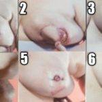
The Nipple stands out in the diagram, surrounded by its ever-present pigmented neighbor called the areola.
The underlying structures shown in the diagram are the pectoralis muscles (Minor and major), the ribs, and the intercostals where we have intercostal structures like muscles, nerves, and blood vessels. These are parts of the chest wall.
Whereas ‘costal’ refers to the ribs, intercostals mean ‘between the ribs’. In a young lady, the nipples are noted to align with the horizontal level of the 4th intercostal. It begins to drop as age and motherhood come into play.
There are also groups of nerves and blood vessels in the breasts as shown in the image.
Before we talk about the mammogram of breast cancer, it will help us if we first highlight the features of a normal mammogram.
Features of a normal mammogram.
Normally, the appearance or color of any given tissue in a mammogram is density-dependent. Less dense tissues appear as grey while the denser ones appear as white.
Fat or adipose tissues which usually make up much of the breasts are not dense so you would expect much of a normal mammogram to be greyish in appearance. You would also expect some white areas resulting from dense tissues in some women.
The more there are dense tissues, the whiter the mammogram will appear. These are normally healthy dense tissues in a quite normal mammogram.
However, cancerous tumors and a few other abnormalities also appear white.
This is a reason why dense breasts pose a problem in the detection of breast cancer apart from the fact women with denser breasts are at a higher risk of developing breast cancer.
The denser the breasts, the more challenging it will be to detect cancer cells or a few other abnormalities we will discuss later in this article.
So based on breast density, four types of mammograms are considered normal. These are aligned with the categories of breast densities. We will discuss breast density in a later article.
Meanwhile, you will find further information with typical images by the American Cancer Society (ACS).
Be informed that breast density is part of a mammogram report or findings and not what you can tell by the touch or feeling of the breasts.
Features of an abnormal mammogram.
there are a few findings that could feature in a mammogram that make it abnormal. Apart from cancerous lumps, others are:
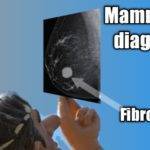
Fibroadenomas:

It is common among ladies below the age of 36 years but is not age-restricted.
Breast Calcifications:

Image credit: Radiology Assistant, Netherlands
Depending on the size, they could be described as micro or macrocalcifications.
Scar tissues:
It is common knowledge these are dense tissues so they also appear white in a breast mammogram. Anyone that has scar tissue will usually know by the feel so it is expedient you inform your doctor about it before your mammogram so it is not mistaken for something else.
Cysts:

They are usually non-cancerous.
Mammogram of breast cancer:
Breast mammogram screenings are aimed at revealing cancerous growths before they become symptomatic. When these cancerous or malignant cells are detected early in mammograms, they are usually small and yet to spread beyond the breasts.
Early detection of these cancerous cells generally has a better prognosis. That is, they help in the achievement of good treatment outcomes.
Note that Initial observations on a mammogram are only indications of abnormalities rather than the definite diagnosis of breast cancer.
Areas of high-density lumps are the targets and their observable shapes, sizes, and edges are evaluated.
According to a publication in the National Center for Biotechnology Information,(NCBI) these appearances really matter in pathology considerations.2

Though they are not the only tissues with that color characteristic as highlighted earlier, further tests and comparative studies will reveal which is malignant(cancerous) and which is not.

Having seen a mammogram of breast cancer, let us compare it with a mammogram of an extremely dense breast. This is for you to understand why it is challenging to detect cancer in a dense breast and the reason you must seek to get screened by the best hands in the industry.

A study published in the Journal of American College of Surgeons has shown that breast cancers in women younger than 36 years of age usually present with quite distinct and palpable mass rather than just mammographic detection.3
The prognosis (future expectation following medical intervention) of breast cancer in these younger women is poorer than is obtainable in older women. Additionally, this class of ladies is more likely to undergo a mastectomy than older women.
I know, at the mention of mastectomy, one thing that comes to the mind of most ladies is the impact of this surgical procedure on their sex life and relationships.
Well, the American Cancer Society (ACS) once again, has a surprise for everybody.4
A study conducted by the society revealed that a woman’s sex life before the advent of cancer has a greater influence on her post-treatment sex life, much more than any resultant damages to her breast from surgery. Share on X
Mammogram report:

This system was developed to ensure uniformity of all mammogram reports across states or regions.
Rate of occurrence of breast cancer in parts of the breast.
This is a graphic image of the rate of occurrence of breast cancer in parts of the breast in relation to others. This research was conducted by Cancer Research, UK.
Some of these parts are anatomically distinct (axillary tail, nipple, and areola ) while others are imaginary(That is, the quadrants).
There are many ways we describe the position of parts of the body In relation to others. For example, we use terms like medial, lateral, posterolateral, superficial, etc.
The quadrants are part of it and imagining it, for instance, helps to easily identify an area for intramuscular injection on the buttocks.

The Axillary tail of the breast accounts for just 1% of all breast cancers, and so is the combination of the nipple and areola.
More than half of all breast cancers (52%) are not specific to any particular location but the vast majority of location-specific cancers occur in the upper outer(lateral) quadrant (25%).
The upper-inner quadrant is the second of all the quadrants with a figure of 8%.
The lower-outer quadrant and lower-inner quadrants account for 5% and 4% respectively.
The last 4% incidence happens at a more central portion of the breast, behind the Nipple.
When you notice anything unusual in your breasts and talk to your doctor about it, the physician will grasp the area you are referring to at the speed of light should you mention the quadrant, for instance.
Congrats, you have just impressed your doctor. However, your moment of heroism will be short-lived should your doctor resort to the use of medical jargon from that point in the belief you will understand.
I hope this article will help you to better understand your mammogram and tell a normal one from an abnormal one. If there are other areas you would like me to discuss do not hesitate to reach out.
- Kothari, C., Diorio, C., & Durocher, F. (2020). The Importance of Breast Adipose Tissue in Breast Cancer. International Journal of Molecular Sciences, 21(16). https://doi.org/10.3390/ijms21165760 ↩︎
- Gajdos, C., Tartter, P. I., Bleiweiss, I. J., Hermann, G., Estabrook, A., & Rademaker, A. W. (2002). Mammographic Appearance of Nonpalpable Breast Cancer Reflects Pathologic Characteristics. Annals of Surgery, 235(2), 246-251. https://doi.org/10.1097/00000658-200202000-00013 ↩︎
- Gajdos, C., Tartter, P. I., Bleiweiss, I. J., Bodian, C., & Brower, S. T. (2000). Stage 0 to stage III breast cancer in young women. Journal of the American College of Surgeons, 190(5), 523-529. https://doi.org/10.1016/S1072-7515(00)00257-X ↩︎
- Schover, L.R. (1991), The impact of breast cancer on sexuality, body image, and intimate relationships. CA: A Cancer Journal for Clinicians, 41: 112-120. https://doi.org/10.3322/canjclin.41.2.112 ↩︎


